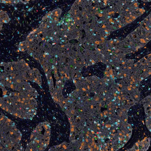
IHC/IF qualitative or quantitative evaluation (‘scoring’)
The interpretation of the tissue staining by different techniques, like immunohistochemistry, immunofluorescence, in situ sequencing, FISH, SISH etc., is challenging and needs knowledge in tissue morphology.
What is stained? Is this staining specific? Is your marker expressed in cancer cells or in stroma cells? Which regions are necrotic? Are there artefacts in the image? Or maybe there are small regions of non-malignant tissue present in your tissue sample labelled as 'tumour'?
A reliable scoring is thus only possible based on good histopathology expertise.
We can provide professional interpretation, evaluation and quantification of staining, considering tissue complexity and specific needs of your research project.
Completing missing pathological information*
In tissue collections used in the research community, pathological information is often missing. But in many cases, it can be re-constructed, by accessing the tissue used for the analysis. We can complete such information like tumour grading, mitotic count, dysplasia grade, specify lesion sub classification etc for your samples.
*for non-clinical use
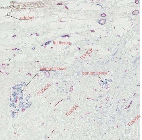
Image curation and quality control
Digital image analysis is more and more broadly used in the research community and multiple applications can be applied to accurately quantify diverse features of the tissue. An often-neglected aspect is the primary composition of the evaluated tissue. Do the samples provide the intended tissue? Diseased tissue samples often contain large areas of necrotic tissue, debris, or even artefacts. Often areas of histological normal tissue might be present within your tissue sample. Such features can be undistinguishable for non-professional and may lead to dramatic impact on the results of image analysis.
We offer image 'curation' (i.e. removal of artefacts, necrosis, irrelevant areas etc) and quality control as a preparation for any further image analysis.
Education in pathology for biomedical researchers
To became a pathologist, you need 6 years of hard training at university and many years afterwards as pathology trainee. However, basic understanding of certain specific features in certain tissue type may be achieved much faster. If you need to distinguish lymphocytes from histiocytes, adenocarcinoma from squamous cell carcinoma, be able to recognize arteries and veins on H&E staining, you can learn it, probably, in few hours. Proper, interactive mentoring in crucial in this process. By demand, we can arrange individual virtual classes for such kind of training.
Slide digitalization
We don’t provide the service for slide digitalization. However, we can assist to arrange slide scanning service vie third parties.